Showing posts with label Lupine Publishers Group. Show all posts

Friday, 10 February 2023

Prevention and Correction of Immunodeficiency States of Animals, Chemical Etiology

 Introduction

In recent years, in farm and private livestock farms in Uzbekistan there has been a decrease in the resistance of animals and, especially, young animals to various bacterial and viral infections. Often there is a decrease and a complete lack of protective action of known vaccines and serums, which causes serious economic damage to livestock. One of the leading factors of this pathology is the unfavorable ecological situation which has developed in many regions of the Republic owing to various anthropogenic influences, including: application of pesticides and other toxic xenobiotics and also emissions of industrial productions.

 Read more about this article: https://lupinepublishers.com/dairy-veterinary-science-journal/fulltext/prevention-and-correction-of-immunodeficiency-states-of-animals-chemical-etiology.ID.000133.php

Read more Lupine Publishers Google Scholar Articles :  https://scholar.google.com/citations?view_op=view_citation&hl=en&user=G7luzkMAAAAJ&citation_for_view=G7luzkMAAAAJ:bFI3QPDXJZMC

Thursday, 1 December 2022

Prevention of Negative Impact of Pesticides and Other Toxic Substances on Reproductive Function of Animals

Introduction


In acutely facing the problem of remote consequences of influence on an organism of chemical substances, impact on reproductive function is important. In recent years, in farms and private farms specializing in the cultivation of cattle and small cattle, as well as rabbits, there are violations of reproductive function, manifested in infertility and spontaneous abortions, pre - and postimplantation developmental delay, physiological and behavioral changes in the offspring, malformations and transplacental effects, and often their etiology remains unclear.


Read more about this article: https://lupinepublishers.com/dairy-veterinary-science-journal/fulltext/prevention-of-negative-impact-of-pesticides-and-other-toxic-substances-on-reproductive-function-of-animals.ID.000134.php

Read more 
Lupine Publishers Google Scholar Articles :   https://scholar.google.com/citations?view_op=view_citation&hl=en&user=G7luzkMAAAAJ&citation_for_view=G7luzkMAAAAJ:bFI3QPDXJZMC

Friday, 25 February 2022

Lupine Publishers | Waste Generated by Food Industry and Reuse in A Circular Economy Approach: The Whey Processing

  Lupine Publishers | Journal of Diary & Veterinary sciences


Mini Review

The popularity of the circular economy is due to the increasing amount of waste-produced in the agro-food processing industry; new solution of waste recycling with biotech innovation are available. In the EU 3.5 ton per capita of waste are annually produced, including more than 400kg per person per year of domestic waste. The projections suggest that this increase at worldwide level, will continue at least until 2030 and there is no real evidence of decoupling between waste and economic growth despite progresses in waste recycling. While all sectors are potentially eligible for funding under the Eco-innovation initiative, certain activities have been singled out as priority areas because of their considerable impact on the environment and their potential contribution to meeting the EU’s own environmental objective. In the modern Agro-food system, the proper treatment of organic effluents to avoid their discharge as sewage water or sewage sludge, to prevent the pollution of the ground and water resources (oceans, lakes, rivers) is becoming especially important. Water is essential not only for direct uses, but also for ensuring the integrity of the ecosystems and the goods and services they provide to humans. The case we have considered is the whey, a by product of the cheese production, requiring urgent solutions to improve water efficiency and water quality used in the cycle.

The Cheese Manufacturing and Whey Processing

Cheese whey (CW) is the liquid part after milk has been curdled and strained in cheese production; it is the main by-product of the cheese making [1] After coagulation casein curd separates from the milk, under the action of chymosin or mineral/organic acid producing; the remain is the whey, a watery and thin liquid solution. Approximately from ten parts of milk, one part of cheese and nine parts of whey are produced with appreciable quantity of water soluble components [2]. It is estimated that the whey produced annually by the European dairy industry is about 75 million tons. it is a by-product of cheese making process, in the past it was discharged as waste into soil, rivers, lakes, causing pollution. When poured into a waterway, or sewer, the whey can deplete the water oxygen levels, causing serious environmental damage. The whey pollution is measured by the BOD and COD indexes (1.18) many authors have reported the following results: BOD5 varies between 30 and 60 thousand ppm (35-45 kg/m3) while COD varies between 50 and 100 thousand ppm.(50-100Kg/m3 ). According to Siso, only 50% of the total quantity of CW is treated and turned into non polluting products, then the whey wastewater disposal of the whey is becoming a major environmental problem in the world with the production of cheese whey is estimated over 108 tonnes per year. The whey dispersion is now forbidden by recent legislation act. In Italy in 2015, 1.2 million tons of cheese and 9.5 million tons of whey were produced; in most of the northern regions the conversion ratio cheese/milk was around 1.1 to 10 due to the prevailing medium hard cheese while in the south the ratio around 1.4 to 10 for the prevailing production of mozzarella cheeses. The hard and semihard cheeses represent the 59% of Italian production, followed by fresh and soft cheeses, with 41%. Four Italian regions located in the northern regions: Lombardia, Emilia Romagna, Veneto and Piemonte produce almost seven million ton of whey representing the 72% of the total amount. Grana Padano is the most diffused hard cheeses accounting for 22% of total milk output. The first step of whey processing is the separation of the retentate fraction containing proteins from permeate fraction containing lactose; different methods are now available as ultrafiltration, diafiltration, inverse osmosis and nanofiltration. Our interest in Lactose is for its use in production of biopolymer namely PHA group after fermentations. PHA (polyhydroxy-alkanoate) is a collective name for a family of biodegradable intracellular bio-polymers made of chemically similar building blocks. PHB (poly-3-hydroxybutyrate) is the most widespread member of the PHA family produced by a wide range of prokaryotic genera starting from renewable feedstocks. A particular characteristic of PHA is its biocompatibility, making them suitable for medical applications. PHA also has good barrier properties, of interest for food product packaging. For these reasons, applications of PHA are found, for instance, in single-use packaging films, bags, containers, paper coatings, agricultural foils, biodegradable carriers for long-term dosage of compounds like drugs or fertilizers, and medical applications like surgical pins, sutures, wound dressings, bone and blood-vessel replacements. Currently, the industrial production of PHA by fermentation, is still a guess in terms of yield, extraction and economic sustainability as the production cost of plastics from petrochemical product is still more competitive and preferred by industrial companies compared to biopolymer production, however the two costs are converging rapidly. The environmental problems associated with the accumulation of traditional petrol derived plastics, due to the long-term degradation, makes urgent to find convenient bioplastic processing. The PHAs are polymers of carbon and reserve of energy accumulated in the cytoplasm of many bacterial species under particular conditions of excess of carbon availability, while some other factors are limiting (i.e. N, P, S, and other). These polymers can be synthesized in different types of PHA that microorganisms accumulate as insoluble inclusion in their bodies. The production of PHA from cost-effective substrates, such as agro-industry residues and specifically the whey is the interest of many researchers, interested in the dairy chain optimization and sustainability. The whey is a by-product of the cheese production chain; in volume represents the 80-90% of the milk converted into cheese. Sweet skimmed whey is subjected to a concentration step, removing 80% of its water content. A convenient solution is to extract proteins from retentate fraction and sell into separate market outlets. The permeate fraction rich in lactose (45gr/liter) is a carbon source for different metabolic pathways. We concentrate in the lactose fermentation to produce PHA; a number of studies identified many microbial groups able to synthesize these polymers, the most important are the Rastonia group, the Escherichia coli, the Capriovidus Necator. These bacterial species are the most used for industrial application since they associate high productivity and reduced times of PHA accumulation. The PHA accumulation speed is very variable: specific rate 0.15 g/g*h equivalent to 15% yield per hour; 16.8 g/L biomass containing 73% PHA were obtained Koller [3].

Biodegradability

Majority of the strains that are able to degrade PHA belong to different taxa such as Gram-positive and Gram-negative bacteria, Streptomyces and fungi. It has been reported that 39 bacterial strains of the classes Firmicutes and Proteobacteria can degrade PHA, PCL, and PBS, but not PLA. The population of aliphatic polymerdegrading microorganisms in different ecosystems was found to be in the following order: PHA > PCL > PBS > PLA. Microorganisms secrete enzymes that break down the polymer (PHA depolymerize) into its molecular building blocks, called hydroxyacids, which are utilized as a carbon source for growth. While degradation by mesophilic temperatures, microorganisms which are capable of degrading various kinds of polyesters at high temperatures are of interest. A thermos-tolerant Aspergillus sp. was able to degrade 90% of PHA film after five days cultivation at 50 °C. In the 1980s, Imperial Chemical Industries developed poly (3-hydroxybutyrateco3- hydroxyvalerate) obtained via fermentation that was named ‘Biopol’. It was sold under the name ‘Biopol’ and distributed in the U.S. by Monsanto and later Metabolix.

Researchers in industry processing are working on methods with which transgenic crops will be developed that express PHA synthesis routes from bacteria to produce PHA as energy storage in their tissue. Commercial ventures scaling up PHA production using fermentation processes include Telles, USA; Biomer Biotechnology Co., Germany; PHA Industrial, Brazil; Mitsubishi Gas Chemical, Japan; Kaneka, Japan; Biomatera, Italy; Jiangsu Nantian Group, China; Tianan Biologic Material, China; and Lianyi Biotech, China. PHAs is a very versatile precursor of bio-plastic materials that raise the attention of different industrial branches. As the best-known and most simple application, these biopolymers are of interest for packaging purposes, especially in such areas where compostable packaging is wanted, e.g. in the food producing industry. Especially in the field of packaging of easily spoiling food, the high oxygen barrier of PHA films is very beneficial. In addition, bottles for shampoos (Wella, Germany) made of PHAs were commercially available in the past. PHAs can be used for paper coating, production of daily commodity items like razors, diapers, hygiene products, or cups and dishes (Metabolix, USA; BASF, Germany). For these applications, PHAs can be processed by techniques of injection moulding or film blowing using the same equipment as known from the well-established processing of petrochemical plastics. In the medical field, PHAs were already investigated as bone implant materials, for tissue engineering, for in-vivo application as implants, surgical pins, screws, meshes and sutures, and as carrier matrices for controlled drug release. Also the production of highly sophisticated surgical articles such as artificial blood vessels and vein valves, spinal fusion cages, bone marrow scaffolds, and meniscus regeneration devices. Especially the possibility to change the composition of PHA allows the manufacture of materials with tailor-made mechanical properties and a fine-tuned degradation rate under in-vivo conditions.

Economic Caveat

Three main problems have to be afforded to make the bioplastic production feasible:

i. cost of feedstock

ii. cost of downstream process

iii. Industrial cost and optimal scale. The feedstock costs are limited by the need to recycle a great amount of whey in intensive cheese production.

By the way marketing opportunities for whey proteins and lactose are growing and compete with PHA production. The 2nd problem is the optimization of the downstream processing for PHA recovery and refining after cell harvest. As intracellular products, PHAs have to be separated from the surrounding non-PHA cell mass, mainly consisting of proteins, lipids, nucleic acids and special polysaccharides. Here, high input with often highly polluting solvents and enormous energy demand still are the caveat in PHA recovery, compromising the demanding claims of these bio-plastics to be ecologically sound materials. The 3rd problem implies to afford the increasing productivity by designing the optimal engineering plant for the final break-through of these biopolymers on the market. A continuous biotechnological production process is well known as an interesting solution for achieving high productivity, lower costs and constant product quality. Some authors reported high productivities of 1.85g/L h for PHB and a constant and satisfying product quality using Cupriavidus necator strain.

To optimize the entire PHA chain, we suggest the following steps:

a) Optimize the collection whey costs from a basin area of enough size to cover the costs and minimize the environmental cost of transport [4].

b) stabilize the whey quality and improve the efficiency of the whey processing through advanced membrane methods of ultrafiltration, nanofiltration, inverse osmosis.

c) Find new bacterial strain to convert directly and more efficiently the lactose into PHA, avoiding the feast-famine two step fermentation.

d) optimize the scale of the chain. Some industries achieved the break point of biopolymer cost production with traditional plastic derived from petroleum (around 1.5 /Kg) with scale production of 40 thousand ton per year [5-7].

 Read More Lupine Publishers diary & veterinary sciences Journal Articles: https://lupine-publishers-dairy-veterinary.blogspot.com/

Friday, 13 March 2020

Confounding of Brucellosis and Blue Tongue in Trans Humane Sheep Flock of Tamilnadu and other Herd Health Impact| Lupine Publishers


Abstract

This present study was carried out to assess the prevalence of brucellosis and blue tongue in a trans humane sheep flock of Tamil Nadu, India. This Sheep flock had a history of inconsistent abortion, repeat breeder, poor fertility rate and higher prevalence of still birth. Serum samples were collected from sheep by random sampling. Serum samples were subjected to Rose Bengal Plate agglutination test (RBT) and ELISA. The risk factors like pregnancy, abortion, age and sex were correlated to the Brucella seropositivity. This study also assessed for the presence of Bluetongue in aborted sheep. It was found that ELISA could be the choice of test for testing of Brucellosis (with the percentage of 57.14). Clinically healthy rams were found to be with brucellosis seropositivity and posed infertility to ewes. It was observed that in trans humane flocks. Brucellosis and Blue tongue has a confounding phenomenon for ovine abortions.
Keywords: Sheep; Trans Humane; Inconsistent Abortion; Brucellosis; Blue Tongue; Confounding

Introduction

Brucellosis is a bacterial zoonosis caused by microorganisms belonging to Brucella, a genus of gram-negative bacteria that behave as facultative intracellular pathogens of ruminants, suidae, canids, and several wildlife species (OIE, 2008). B melitensis is the foremost etiological agent of brucellosis in sheep and goats. It is also the main agent responsible for human brucellosis known as Malta fever. Abortion and infertility are the predominant clinical signs in small ruminants. B. melitensis infection in sheep and goats has been neglected for long time, because small ruminant production was considered as a represents generally low-income activity practiced by landless farmers and marginalized communities in the developing countries. For these reasons the trans humane farming systems continue to have served disease challenges and pose major hurdles in, the control and eradication of many infections. The reasons for such high prevalence may also be result of socio cultural factors, which were compounded by the lack of adequate control measures being applied in small ruminant production systems as per the observations of WHO [1]. The Rose Bengal Plate Agglutinations Test (RBT) was developed originally for the diagnosis of bovine brucellosis and despite a scant information on its usage for Sheep and Goat is available, it is also recommended for the screening of B. melitensis infection in small ruminants [2]. In general, indirect ELISA was considered good test for surveillance purposes in which vaccination is no longer used [3]. Infected nonpregnant livestock may not demonstrate clinical signs of infection and this makes the control and prevention more challenging [4]. Bluetongue, which is caused by the Bluetongue virus (BTV) and transmitted by Culicoides spp. Midges, is a major infectious disease of sheep [5,6]. Among the economic losses resulting from BTV infection are abortion and those due to congenital deformities such as hydranencephaly and cerebellar aplasia in calves and lambs [7]. BTV serotypes -10, 11, 13 and 17 are able to cross the placenta and cause fetal infection [8]. Limited information on concurrent Brucellosis and Blue tongue in Sheep, especially trans humane flocks are available. This study investigated and documented it in a trans humane Sheep flock of Tamil Nadu.

Materials and Methods

Study Area and Flock Details

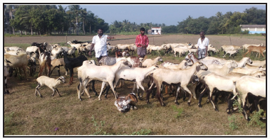
This study was carried out during the period between November 2015 to November 2016 in the Cavery delta districts of Tamil nadu, India. These delta districts are the rice bowl of Tamil nadu state and accounts for 75% of the state’s rice production. The rice harvesting season attracts the several Sheep shepherds to this area to graze the paddy fields after harvesting. Trans humane sheep flocks having flock strength ranging from 300 to 5000 animals per ownership are common sights in these areas. Each flock may have about 300 sheep 25 goat and 9 cattle (Figure 1). These animals were not immunized against brucellosis and blue tongue and no periodical deworming programs are followed. In fact no organized animal health care is being practiced by such nomadic farmers.
Figure 1: Trans human Sheep flock with shepherd in delta districts of Tamilnadu.
Lupinepublishers-openaccess-Dairy-Veterinaryscience

Choice of Samples

Samples were collected from pregnant non aborted, pregnant aborted, non pregnant ewes and rams. For sample size calculation, expected prevalence of brucellosis was assumed as 20 percent with five percent absolute frequency.

Sampling Methods

Clinical materials like whole blood in EDTA tubes, serum collected in clot activator tubes aseptically and stored in refrigeration until further processing. Simple random sampling method was used for sampling.

Brucellosis Screening

Clinical samples like serum samples were collected and submitted for Brucellosis, Blue tongue, Chalymidiasis, Malignant catarrahal fever and Bovine viral diarrhoea diseases since the season and clinical history had given clues for such pathogens. Rose Bengal plate agglutination test (RBT) and I ELISA tests undertaken for brucellosis identification.

Blue Tongue Screening

For blue tongue identification, whole blood in EDTA was collected and subjected to polymerase chain reaction.

Bovine Viral Diarrhoea (BVD) Malignant Cattarhal Fever (MCF) and Chalymidiasis Screening

Serum samples were collected aseptically and analyzed by nested PCR for BVD and MCF. Serology was carried out for Chalymidiasis.

Test Protocols

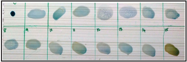
Rose Bengal plate agglutination (RBT) was carried out as per the OIE prescribed procedure. The recommended steps to improve sensitivity of RBT by using three volumes of serum and one volume of antigen (e.g. 75μl and 25μl, respectively) in place of an equal volume of each (Figure 2) This was used in this study modification helped in increasing RBT sensitivity and minimized the discrepancies between RBT and other diagnostic tests. Positive serum and antigen purchased from Indian Veterinary Research Institute (IVRI), Bareilly, India was used in this study. Monoclonal based blocking ELISA for diagnosis of brucellosis was adopted as per the manufactures protocol. For Blue tongue Polymerase chain reaction was adopted as per the standard procedure of OIE.
Figure 2: Modified Rose Bengal plate Agglutination Test.
Lupinepublishers-openaccess-Dairy-Veterinaryscience

Results

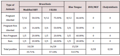
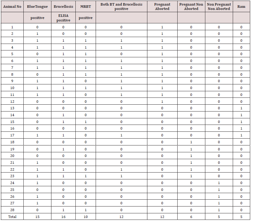
The trans humane sheep population had a clinical history of inconsistent abortion, pooer birth weight of lambs and higher mortality among lambs. Upon screening of 28 clinical samples 15 samples were confirmed as blue tongue with percentage positivity of 53.57. For Brucellosis, out of 10 (35.71%) animals tested by RBT and 16 animals tested by I ELISA (57.14%) were positive for Brucellosis. Out of 12 pregnant aborted animals studied 8 (66.66%) were positive for blue tongue and 7 (58.33%) were shown positive for brucellosis (Table 1) In pregnant non aborted animals, out of the six animals studied three were positive for blue tongue and none of these were positive for brucellosis. Out of five bucks screened, one was positive for blue tongue and two were positive for brucellosis. In non pregnant non aborted animals three and one animals were positive for blue tongue and brucellosis respectively (Table 2). None of the animals were positive for malignant catarrhal fever, Chalymidiasis and Bovine viral diarrhea.
Table 1: Comparison of RBT with ELISA.
Lupinepublishers-openaccess-Dairy-Veterinaryscience
Table 2: Comparison of RBT with ELISA.
Lupinepublishers-openaccess-Dairy-Veterinaryscience

Discussion

Brucellosis and its mode of transmission were known for over 100 years still, the disease remains inconsistent pandemic, predominantly in developing countries. This study analyzed the cause for in consistent abortion in trans- humane sheep flocks of delta regions of Tamil nadu, India. These sheep flocks were nomadic in nature and following flock matting system. They used to exchange their breeding rams with each other over the period of time. The results of the study indicated that 53.57 % of animals were positive for blue tongue and 57.14% were having antibodies for brucellosis which underscored that the incidence was quiet high. Many previous studies had documented it and had an increasing trend [9,10]. The results from this study indicated that brucella antibodies are widely distributed throughout the flock. Since the flock was not immunized in their life time, presence of antibodies directly correlated to the active infection. Prevalence seems to be higher than the overall country seropositivity (13.5%) in Sheep [11-13]. Indirect ELISA showed higher positivity (57.14%) than RBT (35.71%) which indirectly revealed the sensitivity of these tests (Table 3). More over Monoclonal based blocking ELISA for brucellosis detects the antibodies with traces and LPS coated on the ELISA plate has highly homology of the field variant and showed higher titer value (Figures 2 & 3) Conventional Rose Bengal plate agglutination test detects only IgM and is less sensitive, where as ELISA detects both IgM and IgG antibodies which attracts higher sensitivity [2]. Inconsistent abortion was recorded in this flock indicated that it could be the “chronic Brucellosis form”. Rams played vital roles in the transmission of Brucellosis here since higher number (60%) of them showed seropositivity for brucellosis.
Table 3: Categorization of screening tests and positive for Brucellosis and Blue tongue.
Lupinepublishers-openaccess-Dairy-Veterinaryscience
Figure 3: Higher Brucella IELISA titre of Sheep samples.
Lupinepublishers-openaccess-Dairy-Veterinaryscience
In India mostly the sero positive animals were handled based on “test and separate” policy rather than the “test and slaughter” policy due to economic concerns of these marginalized nomadic people. Unplanned immunization program coupled with no effective quarantine and uncontrolled trans-state migration of animals are major factors that affects the Brucellosis control programs [9]. Host susceptibility is also variable with the reproductive status. Thus in the field level, all intermediate stages between typical acute infection to complete resistance may be observed. Under pregnant aborted animals category 58.3% and 66.66% animals showed positivity for Brucellosis and Bluetongue respectively and this highlighted that there is certain confounding effect which makes the animal to abort. In pregnant non aborted category lower percentage of animals showed positive for both Brucellosis and Bluetongue which further supports the possibilities of confounding phenomenon augmenting abortion feature. Interesting by aborted animals got pregnant in their next season and gave birth to normal lambs possibly many animals may develop self limiting infections or they become asymptomatic latent carriers and turn in to potential source of future infections. Abortion generally does not occur if the female is infected at the last stage of pregnancy. Non pregnant non aborted animals showed higher Blue tongue positivity than brucellosis which enlightenend the real risk factor for abortion. Non-pregnant animals exposed to small numbers of organisms may develop selflimiting, immunizing infections or they may become latent carriers [12,13]. No proper selection of breeding ram between flocks of Sheep, low /no biosecurity measures, poor awareness about the vaccination had resulted in brucellosis becoming a continuous threat to the trans humane Sheep populations. As 60 % of rams, showed seropositivity for brucellosis, they posed a great threat to the breeding ewes. Interestingly non of the rams showed clinical signs of Brucellosis. Asymptomatically infected rams deserve to have poor fertility and contributes to dissemination of B. ovis and when very high percentage of rams were infected marked infertility becomes evident in the flock [14]. Seroconversion and shedding of bacteria in the semen of infected rams demonstrated mild or no detectable lesions during the acute phase of infection [15].
The study period of October month had a higher culicids’ populations and it in turn would have helped in transmission of Blue tongue. Results here indicated that BTV is epidemiologically important, and further studies are required to determine the true spatial distribution and cause for abortion in Sheep. Unplanned and uncontrolled grazing and frequent addition of flocks of sheep also contribute to the wide distribution of brucellosis in these animals. Even though the goat and cattle reared along with sheep flock, they were not part of this study, however they may had also acted as a cofounder for Brucellosis. Future studies on these aspects will add more information.


Friday, 28 February 2020

Confounding of Brucellosis and Blue Tongue in Trans Humane Sheep Flock of Tamilnadu and other Herd Health Impact_Lupine Publishers

Abstract

This present study was carried out to assess the prevalence of brucellosis and blue tongue in a trans humane sheep flock of Tamil Nadu, India. This Sheep flock had a history of inconsistent abortion, repeat breeder, poor fertility rate and higher prevalence of still birth. Serum samples were collected from sheep by random sampling. Serum samples were subjected to Rose Bengal Plate agglutination test (RBT) and ELISA. The risk factors like pregnancy, abortion, age and sex were correlated to the Brucella seropositivity. This study also assessed for the presence of Bluetongue in aborted sheep. It was found that ELISA could be the choice of test for testing of Brucellosis (with the percentage of 57.14). Clinically healthy rams were found to be with brucellosis seropositivity and posed infertility to ewes. It was observed that in trans humane flocks. Brucellosis and Blue tongue has a confounding phenomenon for ovine abortions.
Keywords: Sheep; Trans Humane; Inconsistent Abortion; Brucellosis; Blue Tongue; Confounding

Introduction

Brucellosis is a bacterial zoonosis caused by microorganisms belonging to Brucella, a genus of gram-negative bacteria that behave as facultative intracellular pathogens of ruminants, suidae, canids, and several wildlife species (OIE, 2008). B melitensis is the foremost etiological agent of brucellosis in sheep and goats. It is also the main agent responsible for human brucellosis known as Malta fever. Abortion and infertility are the predominant clinical signs in small ruminants. B. melitensis infection in sheep and goats has been neglected for long time, because small ruminant production was considered as a represents generally low-income activity practiced by landless farmers and marginalized communities in the developing countries. For these reasons the trans humane farming systems continue to have served disease challenges and pose major hurdles in, the control and eradication of many infections. The reasons for such high prevalence may also be result of socio cultural factors, which were compounded by the lack of adequate control measures being applied in small ruminant production systems as per the observations of WHO [1]. The Rose Bengal Plate Agglutinations Test (RBT) was developed originally for the diagnosis of bovine brucellosis and despite a scant information on its usage for Sheep and Goat is available, it is also recommended for the screening of B. melitensis infection in small ruminants [2]. In general, indirect ELISA was considered good test for surveillance purposes in which vaccination is no longer used [3]. Infected nonpregnant livestock may not demonstrate clinical signs of infection and this makes the control and prevention more challenging [4]. Bluetongue, which is caused by the Bluetongue virus (BTV) and transmitted by Culicoides spp. Midges, is a major infectious disease of sheep [5,6]. Among the economic losses resulting from BTV infection are abortion and those due to congenital deformities such as hydranencephaly and cerebellar aplasia in calves and lambs [7]. BTV serotypes -10, 11, 13 and 17 are able to cross the placenta and cause fetal infection [8]. Limited information on concurrent Brucellosis and Blue tongue in Sheep, especially trans humane flocks are available. This study investigated and documented it in a trans humane Sheep flock of Tamil Nadu.

Materials and Methods

Study Area and Flock Details

This study was carried out during the period between November 2015 to November 2016 in the Cavery delta districts of Tamil nadu, India. These delta districts are the rice bowl of Tamil nadu state and accounts for 75% of the state’s rice production. The rice harvesting season attracts the several Sheep shepherds to this area to graze the paddy fields after harvesting. Trans humane sheep flocks having flock strength ranging from 300 to 5000 animals per ownership are common sights in these areas. Each flock may have about 300 sheep 25 goat and 9 cattle (Figure 1). These animals were not immunized against brucellosis and blue tongue and no periodical deworming programs are followed. In fact no organized animal health care is being practiced by such nomadic farmers.
Figure 1: Trans human Sheep flock with shepherd in delta districts of Tamilnadu.
Lupinepublishers-openaccess-Dairy-Veterinaryscience

Choice of Samples

Samples were collected from pregnant non aborted, pregnant aborted, non pregnant ewes and rams. For sample size calculation, expected prevalence of brucellosis was assumed as 20 percent with five percent absolute frequency.

Sampling Methods

Clinical materials like whole blood in EDTA tubes, serum collected in clot activator tubes aseptically and stored in refrigeration until further processing. Simple random sampling method was used for sampling.

Brucellosis Screening

Clinical samples like serum samples were collected and submitted for Brucellosis, Blue tongue, Chalymidiasis, Malignant catarrahal fever and Bovine viral diarrhoea diseases since the season and clinical history had given clues for such pathogens. Rose Bengal plate agglutination test (RBT) and I ELISA tests undertaken for brucellosis identification.

Blue Tongue Screening

For blue tongue identification, whole blood in EDTA was collected and subjected to polymerase chain reaction.

Bovine Viral Diarrhoea (BVD) Malignant Cattarhal Fever (MCF) and Chalymidiasis Screening

Serum samples were collected aseptically and analyzed by nested PCR for BVD and MCF. Serology was carried out for Chalymidiasis.

Test Protocols

Rose Bengal plate agglutination (RBT) was carried out as per the OIE prescribed procedure. The recommended steps to improve sensitivity of RBT by using three volumes of serum and one volume of antigen (e.g. 75μl and 25μl, respectively) in place of an equal volume of each (Figure 2) This was used in this study modification helped in increasing RBT sensitivity and minimized the discrepancies between RBT and other diagnostic tests. Positive serum and antigen purchased from Indian Veterinary Research Institute (IVRI), Bareilly, India was used in this study. Monoclonal based blocking ELISA for diagnosis of brucellosis was adopted as per the manufactures protocol. For Blue tongue Polymerase chain reaction was adopted as per the standard procedure of OIE.
Figure 2: Modified Rose Bengal plate Agglutination Test.
Lupinepublishers-openaccess-Dairy-Veterinaryscience

Results

The trans humane sheep population had a clinical history of inconsistent abortion, pooer birth weight of lambs and higher mortality among lambs. Upon screening of 28 clinical samples 15 samples were confirmed as blue tongue with percentage positivity of 53.57. For Brucellosis, out of 10 (35.71%) animals tested by RBT and 16 animals tested by I ELISA (57.14%) were positive for Brucellosis. Out of 12 pregnant aborted animals studied 8 (66.66%) were positive for blue tongue and 7 (58.33%) were shown positive for brucellosis (Table 1) In pregnant non aborted animals, out of the six animals studied three were positive for blue tongue and none of these were positive for brucellosis. Out of five bucks screened, one was positive for blue tongue and two were positive for brucellosis. In non pregnant non aborted animals three and one animals were positive for blue tongue and brucellosis respectively (Table 2). None of the animals were positive for malignant catarrhal fever, Chalymidiasis and Bovine viral diarrhea.
Table 1: Comparison of RBT with ELISA.
Lupinepublishers-openaccess-Dairy-Veterinaryscience
Table 2: Comparison of RBT with ELISA.
Lupinepublishers-openaccess-Dairy-Veterinaryscience

Discussion

Brucellosis and its mode of transmission were known for over 100 years still, the disease remains inconsistent pandemic, predominantly in developing countries. This study analyzed the cause for in consistent abortion in trans- humane sheep flocks of delta regions of Tamil nadu, India. These sheep flocks were nomadic in nature and following flock matting system. They used to exchange their breeding rams with each other over the period of time. The results of the study indicated that 53.57 % of animals were positive for blue tongue and 57.14% were having antibodies for brucellosis which underscored that the incidence was quiet high. Many previous studies had documented it and had an increasing trend [9,10]. The results from this study indicated that brucella antibodies are widely distributed throughout the flock. Since the flock was not immunized in their life time, presence of antibodies directly correlated to the active infection. Prevalence seems to be higher than the overall country seropositivity (13.5%) in Sheep [11-13]. Indirect ELISA showed higher positivity (57.14%) than RBT (35.71%) which indirectly revealed the sensitivity of these tests (Table 3). More over Monoclonal based blocking ELISA for brucellosis detects the antibodies with traces and LPS coated on the ELISA plate has highly homology of the field variant and showed higher titer value (Figures 2 & 3) Conventional Rose Bengal plate agglutination test detects only IgM and is less sensitive, where as ELISA detects both IgM and IgG antibodies which attracts higher sensitivity [2]. Inconsistent abortion was recorded in this flock indicated that it could be the “chronic Brucellosis form”. Rams played vital roles in the transmission of Brucellosis here since higher number (60%) of them showed seropositivity for brucellosis.
Table 3: Categorization of screening tests and positive for Brucellosis and Blue tongue.
Lupinepublishers-openaccess-Dairy-Veterinaryscience
Figure 3: Higher Brucella IELISA titre of Sheep samples.
Lupinepublishers-openaccess-Dairy-Veterinaryscience
In India mostly the sero positive animals were handled based on “test and separate” policy rather than the “test and slaughter” policy due to economic concerns of these marginalized nomadic people. Unplanned immunization program coupled with no effective quarantine and uncontrolled trans-state migration of animals are major factors that affects the Brucellosis control programs [9]. Host susceptibility is also variable with the reproductive status. Thus in the field level, all intermediate stages between typical acute infection to complete resistance may be observed. Under pregnant aborted animals category 58.3% and 66.66% animals showed positivity for Brucellosis and Bluetongue respectively and this highlighted that there is certain confounding effect which makes the animal to abort. In pregnant non aborted category lower percentage of animals showed positive for both Brucellosis and Bluetongue which further supports the possibilities of confounding phenomenon augmenting abortion feature. Interesting by aborted animals got pregnant in their next season and gave birth to normal lambs possibly many animals may develop self limiting infections or they become asymptomatic latent carriers and turn in to potential source of future infections. Abortion generally does not occur if the female is infected at the last stage of pregnancy. Non pregnant non aborted animals showed higher Blue tongue positivity than brucellosis which enlightenend the real risk factor for abortion. Non-pregnant animals exposed to small numbers of organisms may develop selflimiting, immunizing infections or they may become latent carriers [12,13]. No proper selection of breeding ram between flocks of Sheep, low /no biosecurity measures, poor awareness about the vaccination had resulted in brucellosis becoming a continuous threat to the trans humane Sheep populations. As 60 % of rams, showed seropositivity for brucellosis, they posed a great threat to the breeding ewes. Interestingly non of the rams showed clinical signs of Brucellosis. Asymptomatically infected rams deserve to have poor fertility and contributes to dissemination of B. ovis and when very high percentage of rams were infected marked infertility becomes evident in the flock [14]. Seroconversion and shedding of bacteria in the semen of infected rams demonstrated mild or no detectable lesions during the acute phase of infection [15].
The study period of October month had a higher culicids’ populations and it in turn would have helped in transmission of Blue tongue. Results here indicated that BTV is epidemiologically important, and further studies are required to determine the true spatial distribution and cause for abortion in Sheep. Unplanned and uncontrolled grazing and frequent addition of flocks of sheep also contribute to the wide distribution of brucellosis in these animals. Even though the goat and cattle reared along with sheep flock, they were not part of this study, however they may had also acted as a cofounder for Brucellosis. Future studies on these aspects will add more information.


To know more about Open access publishers click on Lupine Publishers

Thursday, 20 February 2020

Occurance of Mycotoxins and Mycotoxicosis in Poultry_Lupine Publishers

Journal of veterinary science| Lupine Publishers

Opinion

Mycotoxins are biologically active, toxic metabolites produced by toxigenic fungi mainly belonging to Aspergillus, Fusarium and Penicillium species, which invade crops in the field and may grow on feedstuffs during storage under favourable conditions of temperature and humidity [1]. FAO estimated that about 25% of food and feedstuffs are contaminated with mycotoxins and strong efforts have been made to decontaminate them by the use of physical and chemical adsorbents but the success made so far is limited [2]. Like other environmental pollutants, mycotoxins also adversely affect the health and productivity in animals and esecially in poultry [3,4]. The economy of poultry industry is heavily affected due to wide mycotoxin exposure or contamination of various agricultural commodities. The economic losses are primarily due to the decreased growth rate, feed conversion efficacy, carcass yield, carcass quality and increased susceptibility to other diseases caused due to their immunosuppressive effects among the affected poultry. A mycotoxicosis is a disease caused by a natural toxin produced by a fungus. In poultry, this usually results when toxin producing fungi grow in grain and feed. Hundreds of mycotoxins have been identified, and many are pathogenic. Mycotoxins may have additive or synergistic effects with other natural toxins, infectious agents, and nutritional deficiencies. Many are chemically stable and maintain toxicity over time. Out of more than 350 mycotoxins identified in nature, aflatoxins, T-2 toxin, diacetoxyscirpenol, vomitoxin, zearalenone, ochratoxins, ergot alkaloids, oosporein, cyclopiazonic acid, and tricothecenes are the most common and important in poultry [5].

Mycotoxicosis and Their Effect in Poultry

Aflatoxicosis: The aflatoxins are toxic and carcinogenic metabolites such as Aspergillus flavus. Aflatoxicosis in poultry primarily affects the liver but can involve immunologic, digestive, and hematopoietic functions. Aflatoxin can adversely affect weight gain, feed intake, feed conversion ratio, pigmentation, carcass yield, egg production, male and female fertility, and large hatchability problems. Some effects are directly attributable to toxins, whereas others are indirect, such as reduced feed intake. Susceptibility to aflatoxins varies, but in general, ducklings, turkeys, and pheasants are susceptible, while chickens and Japanese quail are relatively resistant. Clinical signs vary from general unthriftiness to high morbidity and mortality. At necropsy the lesions are found mainly in the liver, which can be due to necrosis and congestion or yellow due to lipid accumulation. Hemorrhages may occur in liver and other tissues. In chronic aflatoxicosis, the liver becomes yellow to gray and atrophied.
Fusariotoxicosis: The genus Fusarium produces many mycotoxins injurious to poultry. The trichothecene mycotoxins produce caustic and radiomimetic patterns of disease exemplified by T-2 toxin and diacetoxyscirpenol (DAS). Deoxynivalenol (vomitoxin, DON) and zearalenone are common trichothecene mycotoxins that are relatively nontoxic for poultry but may cause disease in large concetratin in feed. Fusariotoxicosis in poultry caused by the trichothecenes results in feed refusal, caustic injury of the oral mucosa and areas of the skin in contact with the mold,acute digestive disease, and injury to the bone marrow and immune system [3]. Lesions include necrosis and ulceration of the oral mucosa, gastro intestinal mucosa, mottling of the liver, atrophy of the spleen and other lymphoid organs, and visceral hemorrhages. In laying hens, decreased egg production can be accompanied by depression, recumbency, feed refusal, and cyanosis evident in the comb and wattles [6]. Other Fusarium mycotoxins cause defective growth of long bones.
Ochratoxicosis: Ochratoxins are quite toxic to poultry. These nephrotoxins are produced chiefly by Penicillium viridicatum and Aspergillus ochraceus in grains and feed. Ochratoxicosis causes primarily renal disease but also affects the liver, immune system, and bone marrow. Severe intoxication causes reduced spontaneous activity, huddling, hypothermia, diarrhea, rapid weight loss, and death. Moderate intoxication impairs weight gain, feed conversion ratio, pigmentation, carcass yield, egg production, fertility, and hatchability [5, 6].
Ergotism: Toxic ergot alkaloids are produced by Claviceps spp, which are fungi that attack cereal grains. The mycotoxins form in the sclerotium, a visible, hard, dark mass of mycelium that displaces the grain tissue. Within the sclerotium are the ergot alkaloids, which affect the nervous system, causing convulsive and sensory neurologic disorders, the vascular system, causing vasoconstriction and gangrene of the extremities and the endocrine system, including neuroendocrine control of the anterior pituitary gland. In chicks, the toes become discolored due to vasoconstriction and ischemia. In older poultry, vasoconstriction affects the comb, wattles, face, and eyelids, which become atrophied and disfigured. Vesicles and ulcers develop on the shanks of the legs and on the tops and sides of the toes. In laying hens, feed consumption and egg production are reduced [6].

Mycotoxicosis Diagnosis in Poultry

Mycotoxicosis should be suspected when the history, signs, and lesions are suggestive of feed intoxication, and especially when moldy ingredients or feed are evident. Toxin exposure associated with consumption of a new batch of feed may result in subclinical or transient disease. Chronic or intermittent exposure can occur in regions where grain and feed ingredients are of poor quality or when feed storage is substandard or prolonged. Impaired production can be a clue to a mycotoxin problem, as can improvement because of correction of feed management deficiencies. Definitive diagnosis involves detection and quantitation of the specific toxins. This can be difficult because of the rapid and high volume use of feed and ingredients in poultry operations. Diagnostic laboratories differ in their respective capabilities to test for mycotoxins and should be contacted before sending samples. Feed and also poultry that are showing sings of sicknes or recently dead should be submitted for pathological examinatios. A necropsy and related diagnostic tests should accompany feed analysis if mycotoxicosis is suspected. Concurrent diseases can adversely affect production and should be considered. Sometimes, a mycotoxicosis is suspected but not confirmed by feed analysis. In these situations, a complete laboratory evaluation can exclude other significant diseases. Feed and ingredient samples should be properly collected and promptly submitted for analysis. Mycotoxin formation can be localized in a batch of feed or grain. Multiple samples taken from different sites increase the likelihood of confirming a mycotoxin formation zone. Samples should be collected at sites of ingredient storage, feed manufacture and transport, feed bins, and feeders [5, 6].

Prevention of Mycotoxicosis in Poultry

Prevention of mycotoxicoses should focus on using feed and ingredients free of mycotoxins and on management practices that prevent mold growth and mycotoxin formation during feed transport and storage. Regular inspection of feed storage and feeding systems can identify flow problems, which allow residual feed and enhance fungal activity and mycotoxin formation. Mycotoxins can form in decayed, crusted feed in feeders, feed mills, and storage bins, cleaning and correcting the problem can have immediate benefits. Temperature extremes cause moisture condensation and migration in bins and promote mycotoxin formation. Ventilation of poultry houses to avoid high relative humidity also decreases the moisture available for fungal growth and toxin formation in the feed. Antifungal agents added to feeds to prevent fungal growth have no effect on toxin already formed but may be cost effective in conjunction with other feed management practices [6]. Propionic acid are effective inhibitor, but the effectiveness may be reduced by the particle size of feed ingredients and the buffering effect of certain ingredients. Sorbent compounds such as hydrated sodium calcium aluminosilicate effectively bind and prevent absorption of aflatoxin. Esterified glucomannan, derived from the cell wall of the yeast Saccharomyces cerevisiae, is protective against aflatoxin B1 and ochratoxins. It reduces toxicity through the binding and reduction in bioavailability of fumonisins, zearalenone, and T-2 toxin. Various other fermentation products, algae and plant extracts, and microbial feed additives have demonstrated ability to bind or degrade mycotoxins and may be applicable and appropriate for the situation.

Acknowledgement

The paper is a part of the research work on the project III 46012 financed by the Ministry of Education, Science and Technological Development of the Republic of Serbia.


To know more about Open access publishers click on Lupine Publishers

Friday, 7 February 2020

Occurance of Mycotoxins and Mycotoxicosis in Poultry| Lupine Publishers

Dairy and Veterinary Sciences| Lupine Publishers

Opinion

Mycotoxins are biologically active, toxic metabolites produced by toxigenic fungi mainly belonging to Aspergillus, Fusarium and Penicillium species, which invade crops in the field and may grow on feedstuffs during storage under favourable conditions of temperature and humidity [1]. FAO estimated that about 25% of food and feedstuffs are contaminated with mycotoxins and strong efforts have been made to decontaminate them by the use of physical and chemical adsorbents but the success made so far is limited [2]. Like other environmental pollutants, mycotoxins also adversely affect the health and productivity in animals and esecially in poultry [3,4]. The economy of poultry industry is heavily affected due to wide mycotoxin exposure or contamination of various agricultural commodities. The economic losses are primarily due to the decreased growth rate, feed conversion efficacy, carcass yield, carcass quality and increased susceptibility to other diseases caused due to their immunosuppressive effects among the affected poultry. A mycotoxicosis is a disease caused by a natural toxin produced by a fungus. In poultry, this usually results when toxin producing fungi grow in grain and feed. Hundreds of mycotoxins have been identified, and many are pathogenic. Mycotoxins may have additive or synergistic effects with other natural toxins, infectious agents, and nutritional deficiencies. Many are chemically stable and maintain toxicity over time. Out of more than 350 mycotoxins identified in nature, aflatoxins, T-2 toxin, diacetoxyscirpenol, vomitoxin, zearalenone, ochratoxins, ergot alkaloids, oosporein, cyclopiazonic acid, and tricothecenes are the most common and important in poultry [5].
Mycotoxicosis and Their Effect in Poultry
Aflatoxicosis: The aflatoxins are toxic and carcinogenic metabolites such as Aspergillus flavus. Aflatoxicosis in poultry primarily affects the liver but can involve immunologic, digestive, and hematopoietic functions. Aflatoxin can adversely affect weight gain, feed intake, feed conversion ratio, pigmentation, carcass yield, egg production, male and female fertility, and large hatchability problems. Some effects are directly attributable to toxins, whereas others are indirect, such as reduced feed intake. Susceptibility to aflatoxins varies, but in general, ducklings, turkeys, and pheasants are susceptible, while chickens and Japanese quail are relatively resistant. Clinical signs vary from general unthriftiness to high morbidity and mortality. At necropsy the lesions are found mainly in the liver, which can be due to necrosis and congestion or yellow due to lipid accumulation. Hemorrhages may occur in liver and other tissues. In chronic aflatoxicosis, the liver becomes yellow to gray and atrophied.
Fusariotoxicosis: The genus Fusarium produces many mycotoxins injurious to poultry. The trichothecene mycotoxins produce caustic and radiomimetic patterns of disease exemplified by T-2 toxin and diacetoxyscirpenol (DAS). Deoxynivalenol (vomitoxin, DON) and zearalenone are common trichothecene mycotoxins that are relatively nontoxic for poultry but may cause disease in large concetratin in feed. Fusariotoxicosis in poultry caused by the trichothecenes results in feed refusal, caustic injury of the oral mucosa and areas of the skin in contact with the mold,acute digestive disease, and injury to the bone marrow and immune system [3]. Lesions include necrosis and ulceration of the oral mucosa, gastro intestinal mucosa, mottling of the liver, atrophy of the spleen and other lymphoid organs, and visceral hemorrhages. In laying hens, decreased egg production can be accompanied by depression, recumbency, feed refusal, and cyanosis evident in the comb and wattles [6]. Other Fusarium mycotoxins cause defective growth of long bones.
Ochratoxicosis: Ochratoxins are quite toxic to poultry. These nephrotoxins are produced chiefly by Penicillium viridicatum and Aspergillus ochraceus in grains and feed. Ochratoxicosis causes primarily renal disease but also affects the liver, immune system, and bone marrow. Severe intoxication causes reduced spontaneous activity, huddling, hypothermia, diarrhea, rapid weight loss, and death. Moderate intoxication impairs weight gain, feed conversion ratio, pigmentation, carcass yield, egg production, fertility, and hatchability [5, 6].
Ergotism: Toxic ergot alkaloids are produced by Claviceps spp, which are fungi that attack cereal grains. The mycotoxins form in the sclerotium, a visible, hard, dark mass of mycelium that displaces the grain tissue. Within the sclerotium are the ergot alkaloids, which affect the nervous system, causing convulsive and sensory neurologic disorders, the vascular system, causing vasoconstriction and gangrene of the extremities and the endocrine system, including neuroendocrine control of the anterior pituitary gland. In chicks, the toes become discolored due to vasoconstriction and ischemia. In older poultry, vasoconstriction affects the comb, wattles, face, and eyelids, which become atrophied and disfigured. Vesicles and ulcers develop on the shanks of the legs and on the tops and sides of the toes. In laying hens, feed consumption and egg production are reduced [6].
Mycotoxicosis Diagnosis in Poultry
Mycotoxicosis should be suspected when the history, signs, and lesions are suggestive of feed intoxication, and especially when moldy ingredients or feed are evident. Toxin exposure associated with consumption of a new batch of feed may result in subclinical or transient disease. Chronic or intermittent exposure can occur in regions where grain and feed ingredients are of poor quality or when feed storage is substandard or prolonged. Impaired production can be a clue to a mycotoxin problem, as can improvement because of correction of feed management deficiencies. Definitive diagnosis involves detection and quantitation of the specific toxins. This can be difficult because of the rapid and high volume use of feed and ingredients in poultry operations. Diagnostic laboratories differ in their respective capabilities to test for mycotoxins and should be contacted before sending samples. Feed and also poultry that are showing sings of sicknes or recently dead should be submitted for pathological examinatios. A necropsy and related diagnostic tests should accompany feed analysis if mycotoxicosis is suspected. Concurrent diseases can adversely affect production and should be considered. Sometimes, a mycotoxicosis is suspected but not confirmed by feed analysis. In these situations, a complete laboratory evaluation can exclude other significant diseases. Feed and ingredient samples should be properly collected and promptly submitted for analysis. Mycotoxin formation can be localized in a batch of feed or grain. Multiple samples taken from different sites increase the likelihood of confirming a mycotoxin formation zone. Samples should be collected at sites of ingredient storage, feed manufacture and transport, feed bins, and feeders [5, 6].
Prevention of Mycotoxicosis in Poultry
Prevention of mycotoxicoses should focus on using feed and ingredients free of mycotoxins and on management practices that prevent mold growth and mycotoxin formation during feed transport and storage. Regular inspection of feed storage and feeding systems can identify flow problems, which allow residual feed and enhance fungal activity and mycotoxin formation. Mycotoxins can form in decayed, crusted feed in feeders, feed mills, and storage bins, cleaning and correcting the problem can have immediate benefits. Temperature extremes cause moisture condensation and migration in bins and promote mycotoxin formation. Ventilation of poultry houses to avoid high relative humidity also decreases the moisture available for fungal growth and toxin formation in the feed. Antifungal agents added to feeds to prevent fungal growth have no effect on toxin already formed but may be cost effective in conjunction with other feed management practices [6]. Propionic acid are effective inhibitor, but the effectiveness may be reduced by the particle size of feed ingredients and the buffering effect of certain ingredients. Sorbent compounds such as hydrated sodium calcium aluminosilicate effectively bind and prevent absorption of aflatoxin. Esterified glucomannan, derived from the cell wall of the yeast Saccharomyces cerevisiae, is protective against aflatoxin B1 and ochratoxins. It reduces toxicity through the binding and reduction in bioavailability of fumonisins, zearalenone, and T-2 toxin. Various other fermentation products, algae and plant extracts, and microbial feed additives have demonstrated ability to bind or degrade mycotoxins and may be applicable and appropriate for the situation.

Acknowledgement

The paper is a part of the research work on the project III 46012 financed by the Ministry of Education, Science and Technological Development of the Republic of Serbia.


Ultrasonic Debridement with Stem Cell Therapy of Suspensory Branch Desmitis in an Equine Patient

Abstract Ultrasonic debridement as a treatment for tendinopathy and desmitis is a relatively new approach in orthopedic surgery. Previousl...